Introduction
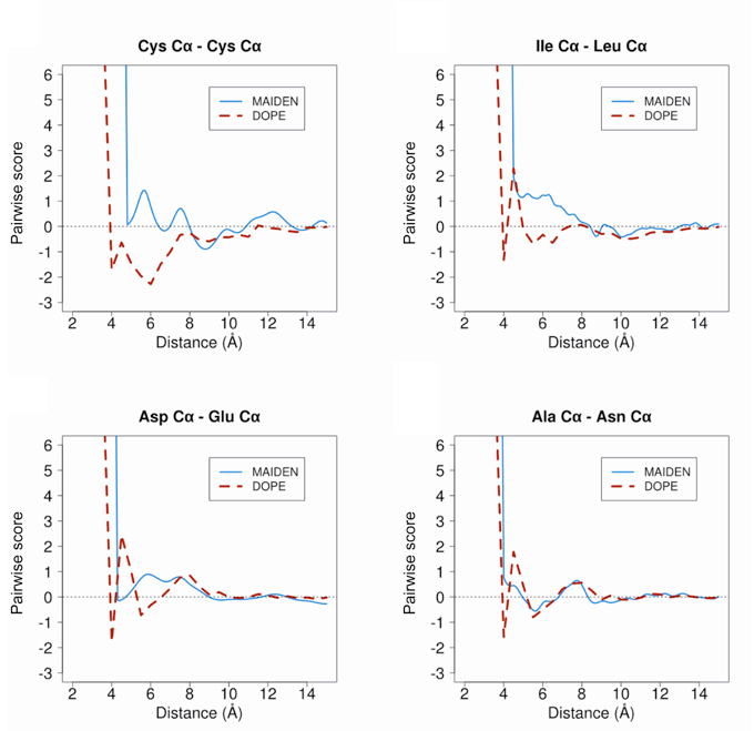
MAIDEN (Model quality Assessment for Intramembrane Domains using an ENergy criterion) is a statistical potential optimized on native alpha-helica and beta-sheet membrane protein structures. MAIDEN computes pseudo-energies for transmembrane domains of native or predicted protein structures, in order to evaluate their quality.
Reference:
Download MAIDEN
Training set
The set of structures used for the calculation of MAIDEN contains 66 representative structures (41 alpha and 25 beta) from the PDBTM database (Tusnady, et al., 2005) determined by crystallography at <2.5 A resolution and with an R-factor <0.3 (see below):
1JB0	2F93	2VDF	3EMN	3SZV	4AL0
1KQF	2FGQ	2W2E	3GIA	3TDS	4AMJ
1OKC	2GR7	2WJR	3GP6	3TIJ	4DVE
1QD5	2HDI	2WSW	3HD6	3TX3	4DX5
1QJP	2J58	2X27	3KVN	3V5U	4E1S
1U19	2J7A	2X55	3M73	3VY8	4EIY
1UUN	2MPR	2X9K	3N5K	3W54	4ENE
1XKW	2O4V	2XOV	3PCV	3WBN	4EZC
1YC9	2Q67	2YEV	3PGU	3ZUX	4HFI
2A65	2QTS	3ARC	3QRA	4A01	4IKW
2BL2	2R9R	3DH4	3RLF	4AFK	7AHL
These representative structures share no more than 30% sequence identity with each other. The list was culled by entries from 1709 PDBTM structure files, using the PISCES server (Wang and Dunbrack, 2003; Wang and Dunbrack, 2005). Fold conservation in transmembrane regions requires less sequence identity than for water-soluble proteins (Olivella, et al., 2013). Therefore, even when sharing <30% sequence identity, proteins belonging to the same structural family based on (Lomize, et al., 2012) were also filtered, making our training set non-redundant both in terms of sequence identity and structure similarity. The assignments of intramembrane residues were obtained using the TMDET web server (Tusnady, et al., 2005).
Models and detailed results
To benchmark MAIDEN and others methods, we have generated 700 predicted structures (alpha and beta) by homology modelling and used the 15340 membrane protein models of EVfold_membrane (Hopf et al., 2012).
Download models and detailed results
References
- Hopf, T.A., et al. Three-dimensional structures of membrane proteins from genomic sequencing. Cell 2012;149(7):1607-1621.
- Lomize, M.A., et al. OPM database and PPM web server: resources for positioning of proteins in membranes. Nucleic acids research 2012;40(Database issue):D370-376.
- Olivella, M., et al. Relation between sequence and structure in membrane proteins. Bioinformatics 2013;29(13):1589-1592.
- Tusnady, G.E., Dosztanyi, Z. and Simon, I. TMDET: Web server for detecting transmembrane regions of proteins by using their 3D coordinates. Bioinformatics 2005;21:1276-1277.
- Wang, G. and Dunbrack, R.L., Jr. PISCES: recent improvements to a PDB sequence culling server. Nucleic acids research 2005;33(Web Server issue):W94-98.
- Wang, Z., Tegge, A.N. and Cheng, J. Evaluating the absolute quality of a single protein model using structural features and support vector machines. Proteins 2009;75(3):638-647.
- Postic G, Ghouzam Y, Gelly JC. An empirical energy function for structural assessment of protein transmembrane domains. Biochimie. 2015 Aug;115:155-61.